The mammography department is situated on the first floor of the Borders General Hospital. From the main entrance, take either the lift on the right or the stairs on the left just after the WRVS coffee shop. On your right as you come out the lift (left if coming up the stairs) is the out patient department. Go into this department and report to the reception on your right. The mammography service is run in conjunction with the breast clinic. We work to an appointed system; patients are referred from their GPs or outpatient clinics for these types of scans.
Your doctor has referred you to the breast clinic. This may be because you have a breast lump, or because of another symptom such as discomfort or discharge.
At the clinic you will be seen by the consultant breast surgeon, or a member of their clinical team. You might spend time in the radiology or x-ray department where you may have a mammogram or breast ultrasound or both. You might have to have a sample of tissue taken from your breast using a small needle.
How long will I be at the clinic?
We try our best to keep you for as short a time as possible. Each test takes about 15 minutes so you have a little bit of a wait if the clinic is busy. After you have had all your tests you will then see the Surgeon so you should expect to spend a good part of the morning or afternoon in the hospital.
Can I bring a relative or friend to the clinic?
Yes, but friends or relatives are not usually allowed in the x-ray rooms. Space is limited in the waiting areas so if possible only bring one person with you. Please avoid bringing children with you to the clinic as there may not be anyone available to look after your child while you are having an examination.
What tests might I need?
Your breasts will be examined by a doctor trained in breast disease. Your breast will be looked at using x-rays or ultrasound (or both) and finally, some fluid or cells may be taken from an area of your breast using a small needle through the skin.
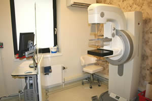
What is a mammogram?
A mammogram is a type of x-ray of the breast. The x-ray is a picture of the insideof the body which is kept in digital form on a computer and looked at on screen.
If you are pregnant, or think you might be, it is important that you tell the radiographer.
You must also tell the radiographer if you have breast implants.
How do I prepare for a mammogram?
It is important that you do not wear talcum powder. Also make sure that there is no deodorant, antiperspirant, body cream or perfume on your breasts.
What happens during the mammogram?
The radiographer will explain the procedure to you and will ask you some questions about your symptoms. You will be asked to undress to the waist and each breast will be x-rayed. The radiographer helps you to place one breast at a time on a small flat plate, with an x-ray plate under it. There is another flat plate above your breast. When the machine is switched on, your breast is pressed down between the plates by the machine for a few moments. This helps to give a clear picture of the breast tissue. It may feel a little uncomfortable but will only last for a short time.
Two x-ray views are taken of each breast from different angles. The radiographer goes behind a screen, but at all times you are in view of the radiographer, and can be heard if you have a problem. You will need to keep still and may hear a light whirring from the x-ray machine.
The radiographer will then check the pictures and if needed repeat part of the process.
You will then be asked to take a seat in the waiting area while your pictures are looked at by the radiologist.
Ultrasound scan of the breast
If you need an ultrasound you will be shown to the radiology department. The Consultant Radiographer or Radiologist will explain the procedure to you.
What is an ultrasound scan of the breast?
Ultrasound scanning uses sound waves to make a picture of the inside of your breast.
You will be asked to undress to the waist and lie on a couch. Some warm gel will be placed on your breast and a small hand held sensor will be pressed against the skin surface. The sensor can be moved over the skin to see the breast from different angles. The lights will be dimmed in the room so that the pictures on the screen can be seen more clearly.
The radiologist or consultant radiographer will be beside you slowly moving the sensor over your skin while looking at the pictures on the screen.
The scan should take around 5-10 minutes.
Fine needle aspiration/core biopsy of the breast
Not everyone who comes to the breast clinic has a fine needle aspiration or core biopsy of the breast.
A fine needle aspiration is a way of taking a few cells from an abnormal area; a core biopsy takes a larger specimen of tissue which can then be looked at under a microscope.
The breast surgeon or the radiologist will do these tests and will explain the procedure to you.
Please tell the Doctor who is doing the test if you are taking any tablets to thin your blood.
After the tests you will be seen by the Consultant breast surgeon who will talk about the results with you.
We aim to make your experience with us as stress free as possible. For any further information or questions regarding these services please contact thedepartment on: 01896 826417 or alternatively complete our online contact form.
<H3>Mammography</H3>
<P style="TEXT-ALIGN: center"><A href="./?a=13024"><FONT size=2>Return to Homepage</FONT></A></P>
<P>The mammography department is situated on the first floor of the Borders General Hospital. From the main entrance, take either the lift on the right or the stairs on the left just after the WRVS coffee shop. On your right as you come out the lift (left if coming up the stairs) is the out patient department. Go into this department and report to the reception on your right. The mammography service is run in conjunction with the breast clinic. We work to an appointed system; patients are referred from their GPs or outpatient clinics for these types of scans. </P>
<P>Your doctor has referred you to the breast clinic. This may be because you have a breast lump, or because of another symptom such as discomfort or discharge.</P>
<P>At the clinic you will be seen by the consultant breast surgeon, or a member of their clinical team. You might spend time in the radiology or x-ray department where you may have a mammogram or breast ultrasound or both. You might have to have a sample of tissue taken from your breast using a small needle.</P> <H4>How long will I be at the clinic?</H4>
<P>We try our best to keep you for as short a time as possible. Each test takes about 15 minutes so you have a little bit of a wait if the clinic is busy. After you have had all your tests you will then see the Surgeon so you should expect to spend a good part of the morning or afternoon in the hospital.</P> <H4>Can I bring a relative or friend to the clinic?</H4>
<P></P>
<P>Yes, but friends or relatives are not usually allowed in the x-ray rooms. Space is limited in the waiting areas so if possible only bring one person with you. Please avoid bringing children with you to the clinic as there may not be anyone available to look after your child while you are having an examination.</P> <H4>What tests might I need?</H4>
<P></P>
<P>Your breasts will be examined by a doctor trained in breast disease. Your breast will be looked at using x-rays or ultrasound (or both) and finally, some fluid or cells may be taken from an area of your breast using a small needle through the skin.</P> <H4>What is a mammogram?</H4>
<P style="TEXT-ALIGN: center"><IMG style="BORDER-BOTTOM: rgb(0,0,0) 0px solid; BORDER-LEFT: rgb(0,0,0) 0px solid; BORDER-TOP: rgb(0,0,0) 0px solid; BORDER-RIGHT: rgb(0,0,0) 0px solid" alt=Mammography src="./?a=13807" width=300 height=200></P>
<P></P>
<P>A mammogram is a type of x-ray of the breast. The x-ray is a picture of the inside of the body which is kept in digital form on a computer and looked at on screen.</P>
<P>If you are pregnant, or think you might be, it is important that you tell the radiographer.</P>
<P>You must also tell the radiographer if you have breast implants.</P> <H4>How do I prepare for a mammogram?</H4>
<P></P>
<P><B>It is important that you do not wear talcum powder. Also make sure that there is no deodorant, antiperspirant, body cream or perfume on your breasts.</B></P>
<P></P> <H4>What happens during the mammogram?</H4>
<P></P>
<P>The radiographer will explain the procedure to you and will ask you some questions about your symptoms. You will be asked to undress to the waist and each breast will be x-rayed. The radiographer helps you to place one breast at a time on a small flat plate, with an x-ray plate under it. There is another flat plate above your breast. When the machine is switched on, your breast is pressed down between the plates by the machine for a few moments. This helps to give a clear picture of the breast tissue. It may feel a little uncomfortable but will only last for a short time.</P>
<P>Two x-ray views are taken of each breast from different angles. The radiographer goes behind a screen, but at all times you are in view of the radiographer, and can be heard if you have a problem. You will need to keep still and may hear a light whirring from the x-ray machine.</P>
<P>The radiographer will then check the pictures and if needed repeat part of the process.</P>
<P>You will then be asked to take a seat in the waiting area while your pictures are looked at by the radiologist.</P> <H4>Ultrasound scan of the breast</H4>
<P></P>
<P>If you need an ultrasound you will be shown to the radiology department. The Consultant Radiographer or Radiologist will explain the procedure to you.</P> <H4>What is an ultrasound scan of the breast?</H4>
<P></P>
<P>Ultrasound scanning uses sound waves to make a picture of the inside of your breast.</P>
<P>You will be asked to undress to the waist and lie on a couch. Some warm gel will be placed on your breast and a small hand held sensor will be pressed against the skin surface. The sensor can be moved over the skin to see the breast from different angles. The lights will be dimmed in the room so that the pictures on the screen can be seen more clearly.</P>
<P>The radiologist or consultant radiographer will be beside you slowly moving the sensor over your skin while looking at the pictures on the screen. </P>
<P>The scan should take around 5-10 minutes.</P>
<P></P>
<P></P> <H4>Fine needle aspiration/core biopsy of the breast</H4>
<P></P>
<P>Not everyone who comes to the breast clinic has a fine needle aspiration or core biopsy of the breast.</P>
<P>A fine needle aspiration is a way of taking a few cells from an abnormal area; a core biopsy takes a larger specimen of tissue which can then be looked at under a microscope.</P>
<P>The breast surgeon or the radiologist will do these tests and will explain the procedure to you.</P>
<P>Please tell the Doctor who is doing the test if you are taking any tablets to thin your blood.</P>
<P>After the tests you will be seen by the Consultant breast surgeon who will talk about the results with you. </P>
<P></P>
<P>We aim to make your experience with us as stress free as possible. For any further information or questions regarding these services please contact the department on: 01896 826417 or alternatively complete our <A href="./?a=13790">online contact form</A>. </P>
<P> </P>